Taming the Zebra – Excerpt 2

Website: tamingthezebra.org

Mailing List: https://www.tamingthezebra.org/join-the-email-list

Excerpt from: Taming the Zebra – It’s Much More than Hypermobility: The Definitive Physical Therapy Guide to Managing HSD/EDS, Volume 1 Systemic Issues and General Approach 

(Due out Winter of 2023)

Fibroblasts and the ECM (Extracellular Matrix)

Those patients with a structurally-compromised ECM – whether from collagen or proteoglycan
abnormalities – often demonstrate the impaired connective tissue function that we see in
connective tissue disorders (Hypermobility Spectrum Disorder, EDS, Marfan’s Syndrome, etc).
Recent research has proposed that different types of EDS can be attributed to different disorders in the ECM. This research has further identified a common cause of fibroblast dysfunction amongst all subtypes of HSD/EDS. Fibroblasts synthesize collagen, create the architecture of the ECM, and play a role in regulating inflammation. So, if fibroblasts do not work properly, neither will the ECM or any system that depends on the ECM.

For example, Malek and Koster (2021) proposes three factors contributing to the connective
tissue dysfunction: receptor interaction, integrin switch abnormalities, and fibroblast dysfunction.

The first issue is a failed interaction between collagen and its receptor, which seems to be
dictated by what type of subgroup of EDS the patient may have. For example, classical EDS is
associated with dysfunction in the type V and I collagen components of the ECM.

The second factor, the integrin switch, is common in other HSD/EDS subtypes. Here, when a
system recognizes the dysfunctional collagen-ECM adhesion, in response, fibroblasts may
compensate by encouraging the cell to adhere to different structures, like fibronectin rather than collagen, as it goes into “survival mode”. This will, unfortunately, feed into the dysfunctional fibroblastic activity.

The third factor concerns dysfunction found within the fibroblast itself. Not only is there an issue
with the fibroblast itself due to the genetic variation suspected, but HSD/hEDS is proposed to
additionally have issues with cell adhesion in the ECM due to the dysfunctional fibroblasts.
Impaired cell-ECM adhesion further impedes the fibroblasts from being able to regulate
homeostasis within their environment (the ECM and connective tissue).

A newer body of research offers that the pathomechanism of the HSD/EDS spectrum as
a whole may be linked to three stages of dysfunction. First, the specific type of EDS or
HSD will dictate the cause of failure between collagen and its receptor. Second, the main
types of HSD/EDS show a response of the integrin switch that recognizes the faulty
collagen-ECM connection and goes into survival mode, binding other ECM ligands
rather than collagen in an attempt to maintain homeostasis and prevent cell death. The
last stage is dysfunction within the fibroblast itself in hEDS, causing abnormal
connective tissue make-up.

Mechano-Sensitivity and Load Tolerance

There are downstream effects on the ECM structure and function due to altered signals when
cells cannot adhere in the usual way or when the structure of collagen is altered. One
downstream issue is increased “mechano-sensitivity”, which is a response to a mechanical
stimulation on the structure.

The ECM in those with EDS has been found to tolerate much lower loads before cells separate
compared to normal ECM. In normal conditions, the ECM responds to loads by becoming
stronger. More research is needed on how the ECM responds to loads in patients with EDS.
Strengthening studies on patients with HSD/EDS report improved strength, pain, proprioception, perceived function of daily living, and stiffness of joints and tendons, indicating that those with HSD/EDS can benefit from strengthening exercises (see Chapter 19). There is a relatively high dropout rate in the studies and we cannot rule out discomfort with the prescribed exercise program as one of the challenges. It is possible that new parameters for strength exercise progression will need to be created that are specific to HSD/EDS, recognizing the difference in ECM response to load. At this point, anecdotal evidence suggests starting with lighter loads/weights and progressing more slowly than standards set out by the American Academy of Sports Medicine with a lower maximum load at the end of the progression to prevent iatrogenic injuries. This may show a more positive response to a strengthening program as there is less load imposed on the ECM with a slower acclimation period, allowing the compromised ECM to adapt more efficiently.
Basics of EDS – Part 2

2022.11.10
Basics, Part 2


Hello, Zeborah Dazzle, PT, WWF here. I am the spokes-zebra and patient educator for Good Health Physical Therapy and Wellness.


As some of you know, while I am a physical therapist who treats all kinds of problems, including all kinds of bone and muscle problems, my special interest is Ehlers-Danlos Syndrome (EDS) and Hypermobility Spectrum Disorder (HSD). Sometimes it is wise to pause and go back to basics. That is what I will focus on with this post.


By the way, in the last post while considering the types of Ehlers-Danlos, I forgot to mention that it occurs across all ethnic and racial backgrounds. And for some patients, inheritance is dominant, meaning that only one parent can pass it to the kids (autosomal dominant) and in some recessive, meaning both parents would have to have the gene to pass it on (autosomal recessive).


What is Hypermobility Spectrum Disorder and How is it Different from Ehlers-Danlos?

In the last post, we talked about how 12 of the 13 types of Ehlers-Danlos Syndrome have genetic tests, but that the most common, hypermobile Ehlers-Danlos Syndrome (hEDS, 80-90% of all EDS) does not. This diagnosis is done by a set of criteria. However, there are many patients who may be generally hypermobile but do not fit the formal criteria for hEDS. These patients are then diagnosed with Hypermobility Spectrum Disorder (HSD), which it should be noted does not mean that they have less pain or fewer symptoms than someone with the diagnosis of hEDS – but more on that later.


At this point in writing this, I see that I have already used the word “hypermobility” many times and used it many times in the last blog post too. So, I think it would be wise to consider what this means.


A joint that is too flexible without adequate soft tissue support is hypermobile.


All joints in the body exist in a balance between flexible and inflexible, or said another way, stable and unstable. Some joints are naturally more stable than others with each joint having its own best level of stability. There are a number of factors which hold the bones together so they can do their job. One factor is the shape of the bones. For example, the bones of the skull are shaped like puzzle pieces which fit together closely. A slight suction between smooth joint surfaces which are lubricated with joint fluid (“synovial fluid”) is another factor which holds bones together — like a suction cup on a windowpane but less strong. By far though, the greatest support to the joints is the soft tissue support around it such as the ligaments and the surrounding muscles. Therapists think of the optimal state of a joint as being flexible and strong.


When a joint is unstable because the soft tissues are broken (such as a severe sprain) or too stretchable or too fragile, this leads to too much mobility at the joint surfaces. This is not just a problem of HSD and hEDS. Even in people with normal connective tissue, sprains, strains and sometimes aging can leave joints too mobile. And weak muscles can also cause local problems to joints. Working with this kind of problem is daily fare for physical therapists. But in HSD and hEDS multiple joints of the body if not all are affected by faulty connective tissue not just one or two localized joints.


So, HSD is an umbrella diagnosis for all patients who are excessively mobile in most or all joints of their body. Like hEDS this is thought to be inherited, but no specific genetic mutation is known. hEDS is under the umbrella of the hypermobility spectrum disorder diagnosis, just more specific.


What does the word “spectrum” mean in Hypermobility Spectrum Disorder?


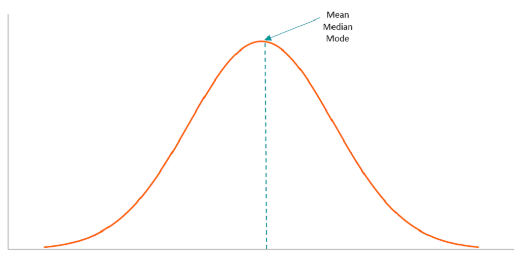
We say that patients with HSD and hEDS are on a spectrum because of the huge variation in symptom levels they can experience. Thinking back to high school, you may remember a thing called a “bell curve”, also sometimes called a normal distribution. This is a graph of how often something happens or how often a specific variable shows up in a data group. With HSD and hEDS, some patients have very few or even no symptoms. In a graph of how many patients with HSD or hEDS have symptoms of different severity, these people would be on the left, mild, side of the curve. Where patients with many or severe problems would be on the right, severe side. Those with moderate problem levels would be the greatest in number and near the average center.

According to the Ehlers-Danlos Society, the occurrence of HSD in the population is about 1/500. This means that out of 332 million people in the US, about 664,000 or .2% are hypermobile. I suspect this estimate is low due to under diagnosing.


I am trying to keep these blog posts bite-sized, meaning about two pages. So, I will continue in the next post and consider the kinds of symptoms that HSD and hEDS can cause, and also what some of the principles of treatment are.


Until then, Cheers! Zebbie


P.S. More information is available at the Ehlers-Danlos Society website. I particularly like this downloadable PDF overview: https://ehlers-danlos.com/wp-content/uploads/EDS_Awareness_2017_v3_img_2021.pdf


Thanks to Dr. Mark Melecki, PT for his assistance in writing this blog. (It is very challenging to type with hooves rather than fingers. Thanks Mark!)