Taming the Zebra Excerpt 3

Website: tamingthezebra.org

Mailing List: https://www.tamingthezebra.org/join-the-email-list

Excerpt from: Taming the Zebra – It’s Much More than Hypermobility: The Definitive Physical Therapy Guide to Managing HSD/EDS, Volume 1 Systemic Issues and General Approach 

(Due out Winter of 2023)

CHAPTER 5

 HSD/EDS and the Skin: Presentation and Management Strategies in Rehab Settings

 As with all EDS presentations, skin involvement varies from person to person and may change in severity throughout one’s life. For this reason, the patient and practitioner need to consider some factors when approaching rehab strategies to avoid exacerbating existing issues. Not all patients with HSD/EDS will present with the issues depicted in this chapter. If a patient or practitioner feels that one of the issues may be present, this is an area to explore individually with appropriate members of the medical team.

 Different classifications of EDS and HSD may have different qualities of skin presentation. Skin hyperextensibility is seen across many different classifications of EDS and more often include cEDS, clEDS, cvEDS, aEDS, dEDS, kEDS, spEDS, mcEDS, pEDS, AEBP1 variant, hEDS, and HSD. Skin fragility (skin that is more easy to tear) is more commonly seen with cEDS, cvEDS, aEDS, dEDS, kEDS-PLOD1, mcEDS, pEDS, AEBP1 variant, and hEDS. Poor wound healing is seen in a variety of severity, more commonly with kEDS-PLOD1, pEDS, AEBP1 variant, and Mast Cell Activation Syndrome (MCAS). Easy bruising is more commonly seen with cEDS, clEDS, cvEDS, vEDS, aEDS, dEDS, kEDS, pEDS, and sometimes with hEDS. Easy bruising that is cyclical (comes and goes) may be more related to systemic flare-ups, such as MCAS reactions.

This chapter presents the different types of skin involvement that might be seen in the clinic and if that issue is present, how to modify treatment or interventions as needed for better outcomes. The individual presentation should always be considered with modifications implemented as needed for the best outcomes in therapy.

 Unusual Skin Presentations

 There are certain skin presentations in HSD/EDS that do not have a major impact on rehabilitation outcomes. For example, the presence of soft, velvety, doughy skin texture does not usually impact therapeutic outcomes but may change manual therapy approaches. Other similar skin presentations are detailed in Figure 5.1.

Skin PresentationDescriptionTypical Location
Molluscoid PseudotumorsAreas of fat tissue herniating through atrophic scarsHigh pressure areas, such as the back of the elbows and front of the knees
Subcutaneous SpheroidsAreas of fat that have lost their blood supply and become firm areas of calcification, moveable under the skinLegs and arms
Piezogenic PapulesSoft, fleshy nodules caused by fat herniating through the dermis (skin layers)Heels and wrists
Hemosiderotic PlaquesDiscolorations of the skin generally found in areas of atrophic scarringShins of those with Classical and Periodontal EDS

Figure 5.1 Common skin presentations, description, and typical location.

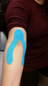
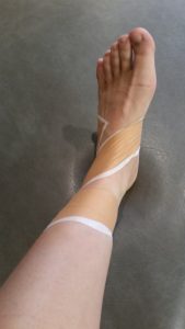
While these skin issues more typically present in HSD/EDS are not necessarily something to treat in the rehab setting, it is important for the therapist to note the level of skin involvement and adjust treatment methods as needed to protect the skin from trauma and tearing, and to avoid tissue scraping over these spots as it will inflame, not improve these lumps and bumps. Many will not be able to tolerate tape without a protective barrier applied first. 

Basics of EDS Part 3

2022.12.16

Hello, Zeborah Dazzle, PT, WWF here. I am the spokes-zebra and patient educator for Good Health Physical Therapy and Wellness. As some of you know, while I am a physical therapist who treats all kinds of problems, including all kinds of bone and muscle problems, my special interest is hypermobile Ehlers-Danlos Syndrome (hEDS) and Hypermobility Spectrum Disorder (HSD). Sometimes it is wise to pause and go back to basics. That is what I will focus on with this post.

What Kinds of Problems Do HSD and EDS Cause?

Both HSD and hEDS are problems with connective tissue. Connective tissue is what holds the body together and it is everywhere in the body. If connective tissue is weak or fragile, many aspects of body function are affected. First in this area is laxity of the skin and joints.

Skin and Joint

Too stretchy connective tissue makes the skin overly stretchy, and also, makes the joints too mobile – hypermobile. The excessive stretchiness of the joints in particular frequently (but not always) leads to pain. When we say ‘not always’ here, remember that in just about every aspect of these conditions, patients fall on a spectrum from few symptoms to severe.

One of the sub-criteria for diagnosis of hEDS in particular is pain in multiple joints lasting more than three months. Mechanically loose joints and weak or overly stretchy connective tissue can lead both to pain and to a number of secondary problems, such as:

early joint damage including spinal disc damage, neck instability, muscle weakness, headaches and migraines, pelvic pain, TMJ (jaw) pain and clicking, gum disease and dental fractures, chiari malformation (connective tissue weakness at the bottom of the skull allows the lower part of the brain to hang out of the skull opening), tethered cord (spinal cord restriction, usually low in the back).

Nervous System

Additionally, HSD and hEDS can affect the nervous system. This can occur in several ways. Loose joints may be less sensitive to what their position is leading to a decrease of the joint awareness (called proprioception) affecting coordination. Prolonged daily pain may cause the nervous system to become sensitized and to create pain sensations  which are out of proportion to the damage being done. And finally, connective tissue deficits can affect the involuntary nervous system of the body.

One of the major divisions of the nervous system is the involuntary, or autonomic, nervous system. The two halves of this system (sympathetic and parasympathetic) are constantly working to keep our bodies in balance and adapted to the world around us. Among the physical functions fully or partially controlled by the involuntary/ autonomic system are: heart rate, breathing rate, blood pressure, opening and closing of the pupils, gut movements for digestion and release of some digestive juices, and temperature regulation.

Research has shown that most people with HSD or hEDS have at least one or more regular symptoms connected to the involuntary/ autonomic nervous system. Sometimes these symptoms are transient, meaning they come and go. Sometimes there are multiple symptoms rising to the level of a problem syndrome. The general term for this is dysautonomia.

Some patients with HSD/hEDS tolerate standing poorly, which may simply be labeled as Orthostatic Intolerance. Some patients may experience chronically low blood pressure caused by the nervous system (Neurally Mediated Hypotension). A common autonomic imbalance affecting patients with is POTS. This is an abbreviation for Postural Orthostatic Tachycardia Syndrome.

The words postural and orthostasis in this refer primarily to the changes in blood flow in the body caused when we change positions such as going from lying to sitting or sitting to standing or just standing for a sustained period. Since the nerves of the involuntary nervous system run through connective tissue, they can be thrown out of balance by flaws or deficits in the connective tissue. So, in POTS, the longer the patient stands, the more blood settles toward the legs and feet, this takes blood away from the brain and from the nerves which sense blood pressure around the heart and in the neck. The drop in blood pressure leads to a fast heart rate (tachycardia) to try to catch up. And the patient becomes faint or dizzy or feels sick. 

Other dysautonomia symptoms may include poor temperature regulation (feeling too hot or too cold), decreased or stalled gut movement in digestion (gastroparesis), and fatigue.

Hernias and Organ Prolapse

Besides the effect of the autonomic nervous system on digestion, connective tissue weakness in the gut can lead to hernias and organ prolapse plus functional problems like irritable bowel syndrome.

Mast Cell Imbalance

One other syndrome which is commonly associated with HSD or hEDS involves a tiny little cell which lives mostly in the connective tissues and which acts as a frontline sentinel of the body sensing wounds, foreign bodies and infections: the mast cell. Like the autonomic nervous system, mast cells can be thrown out of balance by connective tissue problems which can result in Mast Cell Activation Syndrome.

The mast cells produce a number of chemicals, call mediators, the first function of which is to cause inflammation. When the mast cells are out of balance, they become overly sensitive and may release their inflammatory mediators at random times or in response to triggering events, foods or stimuli. This can result in an array of problems which may appear to be weird and unrelated to each other. Here is a partial list of some of the problems which may result:

abdominal pain, bladder pain, brain fog, brittle fingernails, chemical sensitivity, chronic cough, chronic kidney disease,  chronic sinus irritation or infections, constipation, cystitis, diarrhea, dry eyes/ blurred vision, endometriosis, fatigue, gallbladder inflammation, hair loss, headaches,  hearing loss, high blood pressure, hyper or hypothyroid, incontinence, increased vulnerability to infection, insomnia, laryngitis, loss of appetite, lymph node enlargement, nausea, nose bleeds, numbness, painful urination, rashes, Raynaud’s disease, ringing in ears, sore throat, sores, sweats/ chills, temperature sensitivity, tingling, vaginal inflammation

In the next blog entry, we will look at how HSD and hEDS are treated.

Until then, Cheers! Zebbie

P.S. More information is available at the Ehlers-Danlos Society website. I particularly like this downloadable PDF overview: https://ehlers-danlos.com/wp-content/uploads/EDS_Awareness_2017_v3_img_2021.pdf

Thanks to Dr. Mark Melecki, PT for his assistance in writing this blog. (It is very challenging to type with hooves rather than fingers. Thanks Mark.)

Taming the Zebra – Excerpt 2

Website: tamingthezebra.org

Mailing List: https://www.tamingthezebra.org/join-the-email-list

Excerpt from: Taming the Zebra – It’s Much More than Hypermobility: The Definitive Physical Therapy Guide to Managing HSD/EDS, Volume 1 Systemic Issues and General Approach 

(Due out Winter of 2023)

Fibroblasts and the ECM (Extracellular Matrix)

Those patients with a structurally-compromised ECM – whether from collagen or proteoglycan
abnormalities – often demonstrate the impaired connective tissue function that we see in
connective tissue disorders (Hypermobility Spectrum Disorder, EDS, Marfan’s Syndrome, etc).
Recent research has proposed that different types of EDS can be attributed to different disorders in the ECM. This research has further identified a common cause of fibroblast dysfunction amongst all subtypes of HSD/EDS. Fibroblasts synthesize collagen, create the architecture of the ECM, and play a role in regulating inflammation. So, if fibroblasts do not work properly, neither will the ECM or any system that depends on the ECM.

For example, Malek and Koster (2021) proposes three factors contributing to the connective
tissue dysfunction: receptor interaction, integrin switch abnormalities, and fibroblast dysfunction.

The first issue is a failed interaction between collagen and its receptor, which seems to be
dictated by what type of subgroup of EDS the patient may have. For example, classical EDS is
associated with dysfunction in the type V and I collagen components of the ECM.

The second factor, the integrin switch, is common in other HSD/EDS subtypes. Here, when a
system recognizes the dysfunctional collagen-ECM adhesion, in response, fibroblasts may
compensate by encouraging the cell to adhere to different structures, like fibronectin rather than collagen, as it goes into “survival mode”. This will, unfortunately, feed into the dysfunctional fibroblastic activity.

The third factor concerns dysfunction found within the fibroblast itself. Not only is there an issue
with the fibroblast itself due to the genetic variation suspected, but HSD/hEDS is proposed to
additionally have issues with cell adhesion in the ECM due to the dysfunctional fibroblasts.
Impaired cell-ECM adhesion further impedes the fibroblasts from being able to regulate
homeostasis within their environment (the ECM and connective tissue).

A newer body of research offers that the pathomechanism of the HSD/EDS spectrum as
a whole may be linked to three stages of dysfunction. First, the specific type of EDS or
HSD will dictate the cause of failure between collagen and its receptor. Second, the main
types of HSD/EDS show a response of the integrin switch that recognizes the faulty
collagen-ECM connection and goes into survival mode, binding other ECM ligands
rather than collagen in an attempt to maintain homeostasis and prevent cell death. The
last stage is dysfunction within the fibroblast itself in hEDS, causing abnormal
connective tissue make-up.

Mechano-Sensitivity and Load Tolerance

There are downstream effects on the ECM structure and function due to altered signals when
cells cannot adhere in the usual way or when the structure of collagen is altered. One
downstream issue is increased “mechano-sensitivity”, which is a response to a mechanical
stimulation on the structure.

The ECM in those with EDS has been found to tolerate much lower loads before cells separate
compared to normal ECM. In normal conditions, the ECM responds to loads by becoming
stronger. More research is needed on how the ECM responds to loads in patients with EDS.
Strengthening studies on patients with HSD/EDS report improved strength, pain, proprioception, perceived function of daily living, and stiffness of joints and tendons, indicating that those with HSD/EDS can benefit from strengthening exercises (see Chapter 19). There is a relatively high dropout rate in the studies and we cannot rule out discomfort with the prescribed exercise program as one of the challenges. It is possible that new parameters for strength exercise progression will need to be created that are specific to HSD/EDS, recognizing the difference in ECM response to load. At this point, anecdotal evidence suggests starting with lighter loads/weights and progressing more slowly than standards set out by the American Academy of Sports Medicine with a lower maximum load at the end of the progression to prevent iatrogenic injuries. This may show a more positive response to a strengthening program as there is less load imposed on the ECM with a slower acclimation period, allowing the compromised ECM to adapt more efficiently.

Basics of EDS – Part 2

2022.11.10
Basics, Part 2


Hello, Zeborah Dazzle, PT, WWF here. I am the spokes-zebra and patient educator for Good Health Physical Therapy and Wellness.


As some of you know, while I am a physical therapist who treats all kinds of problems, including all kinds of bone and muscle problems, my special interest is Ehlers-Danlos Syndrome (EDS) and Hypermobility Spectrum Disorder (HSD). Sometimes it is wise to pause and go back to basics. That is what I will focus on with this post.


By the way, in the last post while considering the types of Ehlers-Danlos, I forgot to mention that it occurs across all ethnic and racial backgrounds. And for some patients, inheritance is dominant, meaning that only one parent can pass it to the kids (autosomal dominant) and in some recessive, meaning both parents would have to have the gene to pass it on (autosomal recessive).


What is Hypermobility Spectrum Disorder and How is it Different from Ehlers-Danlos?

In the last post, we talked about how 12 of the 13 types of Ehlers-Danlos Syndrome have genetic tests, but that the most common, hypermobile Ehlers-Danlos Syndrome (hEDS, 80-90% of all EDS) does not. This diagnosis is done by a set of criteria. However, there are many patients who may be generally hypermobile but do not fit the formal criteria for hEDS. These patients are then diagnosed with Hypermobility Spectrum Disorder (HSD), which it should be noted does not mean that they have less pain or fewer symptoms than someone with the diagnosis of hEDS – but more on that later.


At this point in writing this, I see that I have already used the word “hypermobility” many times and used it many times in the last blog post too. So, I think it would be wise to consider what this means.


A joint that is too flexible without adequate soft tissue support is hypermobile.


All joints in the body exist in a balance between flexible and inflexible, or said another way, stable and unstable. Some joints are naturally more stable than others with each joint having its own best level of stability. There are a number of factors which hold the bones together so they can do their job. One factor is the shape of the bones. For example, the bones of the skull are shaped like puzzle pieces which fit together closely. A slight suction between smooth joint surfaces which are lubricated with joint fluid (“synovial fluid”) is another factor which holds bones together — like a suction cup on a windowpane but less strong. By far though, the greatest support to the joints is the soft tissue support around it such as the ligaments and the surrounding muscles. Therapists think of the optimal state of a joint as being flexible and strong.


When a joint is unstable because the soft tissues are broken (such as a severe sprain) or too stretchable or too fragile, this leads to too much mobility at the joint surfaces. This is not just a problem of HSD and hEDS. Even in people with normal connective tissue, sprains, strains and sometimes aging can leave joints too mobile. And weak muscles can also cause local problems to joints. Working with this kind of problem is daily fare for physical therapists. But in HSD and hEDS multiple joints of the body if not all are affected by faulty connective tissue not just one or two localized joints.


So, HSD is an umbrella diagnosis for all patients who are excessively mobile in most or all joints of their body. Like hEDS this is thought to be inherited, but no specific genetic mutation is known. hEDS is under the umbrella of the hypermobility spectrum disorder diagnosis, just more specific.


What does the word “spectrum” mean in Hypermobility Spectrum Disorder?


We say that patients with HSD and hEDS are on a spectrum because of the huge variation in symptom levels they can experience. Thinking back to high school, you may remember a thing called a “bell curve”, also sometimes called a normal distribution. This is a graph of how often something happens or how often a specific variable shows up in a data group. With HSD and hEDS, some patients have very few or even no symptoms. In a graph of how many patients with HSD or hEDS have symptoms of different severity, these people would be on the left, mild, side of the curve. Where patients with many or severe problems would be on the right, severe side. Those with moderate problem levels would be the greatest in number and near the average center.

According to the Ehlers-Danlos Society, the occurrence of HSD in the population is about 1/500. This means that out of 332 million people in the US, about 664,000 or .2% are hypermobile. I suspect this estimate is low due to under diagnosing.


I am trying to keep these blog posts bite-sized, meaning about two pages. So, I will continue in the next post and consider the kinds of symptoms that HSD and hEDS can cause, and also what some of the principles of treatment are.


Until then, Cheers! Zebbie


P.S. More information is available at the Ehlers-Danlos Society website. I particularly like this downloadable PDF overview: https://ehlers-danlos.com/wp-content/uploads/EDS_Awareness_2017_v3_img_2021.pdf


Thanks to Dr. Mark Melecki, PT for his assistance in writing this blog. (It is very challenging to type with hooves rather than fingers. Thanks Mark!)

Taming the Zebra

Website: tamingthezebra.org

Mailing List: https://www.tamingthezebra.org/join-the-email-list

Excerpt from: Taming the Zebra – It’s Much More than Hypermobility: The Definitive Physical Therapy Guide to Managing HSD/EDS, Volume 1 Systemic Issues and General Approach 

(Due out Winter of 2023)

CHAPTER 2

 Understanding Connective Tissue

The Ehlers-Danlos Syndromes (EDS) are described as a group of heritable heterogenous connective tissue disorders, meaning different genetic variations are present with different classifications of EDS. EDS is not simply a diagnosis of joint hypermobility, but a reference to a connective tissue disorder throughout the body, involving many different systems. Presentation with each patient will be determined by the type of genetic variation identified along with genetic expression, which is further discussed below.

The human body is made up of nervous, muscular, epithelial (skin), and connective tissue. Connective tissue can be found in the nervous and muscular tissue and adjacent to the epithelial tissue. Connective tissue plays many different roles for us within our bodies (Figure 2.1). It helps package and compartmentalize areas of the body by providing support or protection. It can bind and separate organs or other tissues. Connective tissue also plays a role in protection, defense, and repair. It aids in scar tissue formation, inflammation, and defense against invading bacteria or other substances through some of its molecular components. It acts as insulation, storing energy as adipose tissue (fat). It also assists in transportation throughout the body. Blood is a connective tissue that delivers oxygen and nutrients throughout the body. Blood is considered a connective tissue because it consists of blood cells surrounded by a fluid matrix called blood plasma. Fascia is a connective tissue creating a continuous system throughout the body, becoming a means of directing and transferring mechanical forces within the body. If, however, the connective tissue is dysfunctional, this can lead to the transfer of inefficient forces and lead to imbalances and/or restrictions. It is thought that the connective tissue is the medium for acupuncture treatment and explains how needles affect organs from afar. Myofascial release experts purport that memory can be stored in the guarding patterns of the tissue, explaining some chronic, non-responsive fascial dysfunction. Connective tissue is complex and expansive within the human body.

Roles of Connective Tissues Throughout the Body
Packaging and Compartmentalizing
Protection, Defense, and Repair
Insulation
Transfer of Mechanical Forces Throughout the Body
Figure 2.1 Connective tissues assists with many different functions and roles within the human body. A connective tissue disorder can cause issues in any of these roles listed.

Connective tissue is the most abundant tissue in our body, found just about everywhere. It is found in fibrous tissues, fat, cartilage, bone, bone marrow, tendons, the wall of the gastrointestinal system, skin, and blood vessel walls. It also encloses the brain and spinal column. Connective tissue is made up of many different components, primarily elastin, collagen fibers, ground substance (gelatinous material that fills the spaces between fibers and cells), and immune cells. Those collagen fibers along with proteoglycans (protein) and glycosaminoglycans (polysaccharide compound) together make up the extracellular matrix along with other compounds. The distribution and ratio of each of these in a particular make-up of connective tissue will determine what the connective tissue looks like (i.e. fibrous versus ligamentous). The function of the connective tissue is determined by the protein composition of the extracellular matrix (ECM). The immune cells reside in the extracellular matrix. 

Figure 2.2 Connective tissue within the human body makes up cartilage, tendon, bone, adipose tissue, and ligaments. Connective tissue surrounds the blood vessel walls, muscles, and nerves, also influencing these systems as well.

Basics of EDS

2022.11.10
Basics
Hello, Zeborah Dazzle, PT, WWF here. I am the spokes-zebra and patient educator for Good Health Physical Therapy and Wellness.


As some of you know, while I am a physical therapist who treats all kinds of problems, including all kinds of bone and muscle problems, my special interest is Ehlers-Danlos Syndrome (EDS) and Hypermobility Spectrum Disorder (HSD). Sometimes it is wise to pause and go back to basics. That is what I will focus on with this and the next post.


What is Ehlers-Danlos Syndrome?
Ehlers-Danlos Syndrome (EDS) is a group of inherited connective tissue disorders. At present, there are thirteen variations of the disease. All involve fragility of one or more of the 28 types of collagen which is the major part of connective tissue.


Connective tissue supports, protects and holds the tissues of our bodies together. It includes tendons, ligaments, blood and the support structures for arteries, veins and muscles as well as internal organs. It is very important in the body, and it is EVERYWHERE. A colleague of mine likes to tell patient’s that if he was an evil wizard and waved a wand that took away all other cell types in your body other than connective tissue, there would still be a perfect three dimensional you remaining. (You would be dead though and we could probably shine a flashlight through you – bad wizard.)


Among the thirteen different types of EDS are types which predominantly affect different parts of the body especially the heart, the blood vessels, the eyes, skin, gums and bones/ joints. Here is a list of the names of the thirteen.


How Common is Ehlers-Danlos Syndrome?
The different EDS types range from uncommon to very rare in the general population. The single most common type of EDS is Hypermobile Ehlers-Danlos Syndrome (hEDS). The prevalence of this condition is estimated by the Ehlers-Danlos Society as 1/3500 to 1/5000 people. That means that out of the 332,218,200 people in the US there may be 94, 919 (0.02%) of the population with hEDS. The other kinds of EDS are much less common ranging from 1/40,000 (about 8,305 people, .003%) to 1/1 million people (about 332 people). I believe these estimates are low due to under diagnosing.


How is Ehlers Danlos Diagnosed?
Of the thirteen different types, there are genetic tests for all but the most common, hEDS. As a result, the diagnosis of hEDS is performed by evaluating a patient through three criteria levels. In the first level, hypermobility, or excessive stretch/ flexy/ bendy-ness is screened for. In the second level, physical characteristics commonly associated with hEDS are screened including some physical characteristics, skin texture and stretchiness and common medical history indicators. In the last criteria level, a physician must rule out other conditions which can mimic the symptoms of hEDS. It is common for patients to come to see me and want me to diagnose hEDS. Physical therapists are trained to evaluate and diagnose movement disorders and to correlate these with medical problems which sometimes means screening for problems. So, as a PT, I can diagnose hypermobility but only screen for hEDS. I cannot not diagnose it formally because I cannot rule out other conditions. Patients who are hypermobile but do not fit the diagnostic criteria of hEDS are diagnosed with hypermobility spectrum disorder (HSD). More on this in a later post.


Why Do People with Ehlers-Danlos Syndrome Call Themselves “Zebras”?
In the next post, we will go into more detail about the kinds of problems that EDS can cause, but for now let me simply say that they are multiple and often appear to be unrelated – joint pain in many areas of the body, bruising, strains, sprains, subluxations or dislocations, gut problems, dizziness and many more. For this reason, people with EDS do not fit in to a quick common pattern for diagnosis.
When medical students are trained, they spend a great deal of time learning about different conditions and the symptoms they can create. Because common symptoms can so often be caused by different underlying conditions, this makes diagnosis hard. Almost all medical students are taught this aphorism: If you hear hoofbeats, think horses, not zebras.


This is a good thing overall. Afterall, if Mrs. Smith comes in to see the doctor complaining of a sore throat, the most probable cause is something common. Perhaps she was shouting at a football game or perhaps she has a cold. Throat cancer is much farther down the list.


The problem for EDS patients though is that we are actually zebras (or in my case a zebra-zebra). Beginning medical training in anatomy is changing in medical school these days, but traditionally anatomy was taught by cadaver dissection. During this process, connective tissue is commonly the stuff put in the container under the table while the student is looking for another structure like a nerve or a blood vessel or an organ or a muscle. Is it any surprise, that a connective tissue disorder is often not on the doctors mental list of potential diagnoses? I am hopeful that this is changing, but in the meanwhile, it is up to all of us with HSD or EDS to educate ourselves so we can partner with our providers in the most positive way.


In the next post, we will look more closely at hypermobility spectrum disorder and the symptoms that HSD and EDS can create as well as an overview of treatment principles.


Until then, Cheers! Zebbie


Thanks to Dr. Mark Melecki, PT for his assistance in writing this blog. (It is very challenging to type with hooves rather than fingers. Thanks Mark.)

What is the difference between Kinesio®tape and athletic tape?

Cueing vs stability
Kinesio® tape is stretchy compared to athletic and Leukotape®. Each are great and beneficial, but for very different purposes.

The stretchy quality of Kinesio® tape provides cues for better joint alignment and position, muscle activation, and posture. As your body moves into these positions, the tape will stretch, giving you a cue to reduce the tension of the tape and return to the better posture and alignment. The stretch of the tape can also be used to help lift the skin, allowing more blood flow. It has been shown to help reduce swelling and help with soft tissue healing.

Other kinds of more traditional tape are used for stabilization. Because of its less stretchy nature, athletic tape will prevent movement into poor positions all together, not allowing you to move out of a certain alignment.

How do you know if Kinesio® tape or athletic tape would be best for you?

Kinesio® tape to help prevent elbow hyperextension

Leukotape® to keep the arch lifted and prevent twisting the ankle

The best way to know would be to see a physical therapist to assess what is happening with your posture or joint alignment. We are trained in the different techniques, patterns, and ways to cut and apply the tape to provide the best benefit and proper purpose of the tape.

For a quick answer though, if you are looking for more stability like a temporary brace, then athletic or Leukotape® are best. If you are trying to retrain yourself to use the right muscles and maintain a certain posture or alignment, then Kinesio® tape is best.


We recommend seeing a physical therapist for the other more medical applications of Kinesio® tape such as to reduce swelling or increase blood flow to an injured area for healing.

A physical therapist at Good Health Physical Therapy & Wellness is always happy to provide more information or assess your needs to reduce or prevent pain and injury

Oregon is a direct access state for physical therapy. What does this mean for you?

Many people think that you need a doctor’s referral to get specialized care. People often see their primary care physician first for any ailment, and your doctor directs you to the right specialist for further investigation and diagnosis. Recently, with rising healthcare costs and a change in physical therapy training, many states now allow direct access. This means if you have a condition involving your muscles or joints you have the right to see a physical therapist (a musculoskeletal specialist) without a referral from your doctor. This can depend on your insurance policy, however. While this is not a federal law, several states have some version of direct access. Oregon is one of them!


According to a 2016 article in the American Physical Therapy Association’s magazine, PT in Motion, Oregon is one of several states that provide unrestricted direct access. A study done by the American Physical Therapy Association has shown the same quality of care, no adverse events and lower cost for patients who saw a physical therapist through direct access compared to those who went to a primary care provider first.


Physical therapists are trained to rule in and rule out red flag signs and symptoms. If we are in doubt about a diagnosis, then we refer you back to a medical doctor to receive the appropriate care. More often, though, people see their doctor for a muscle or joint condition and are then referred to a physical therapist. Direct access allows you to skip a step, make one less appointment, save money, and go directly to the person who can treat your symptoms. You wouldn’t go to a physical therapist first for a sore throat, and you shouldn’t go to your primary care provider first for a pulled muscle.


So, if you think you have a musculoskeletal injury, see your physical therapist first! Start by calling our office (503)292-5882 and our helpful staff will get you an appointment. They can check your insurance company’s requirements/coverage or explain more about our reasonable cash pay rates.


Chie Tadaki, PT, DPT